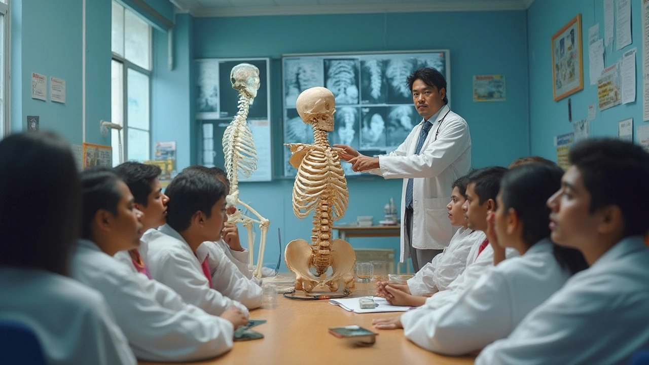
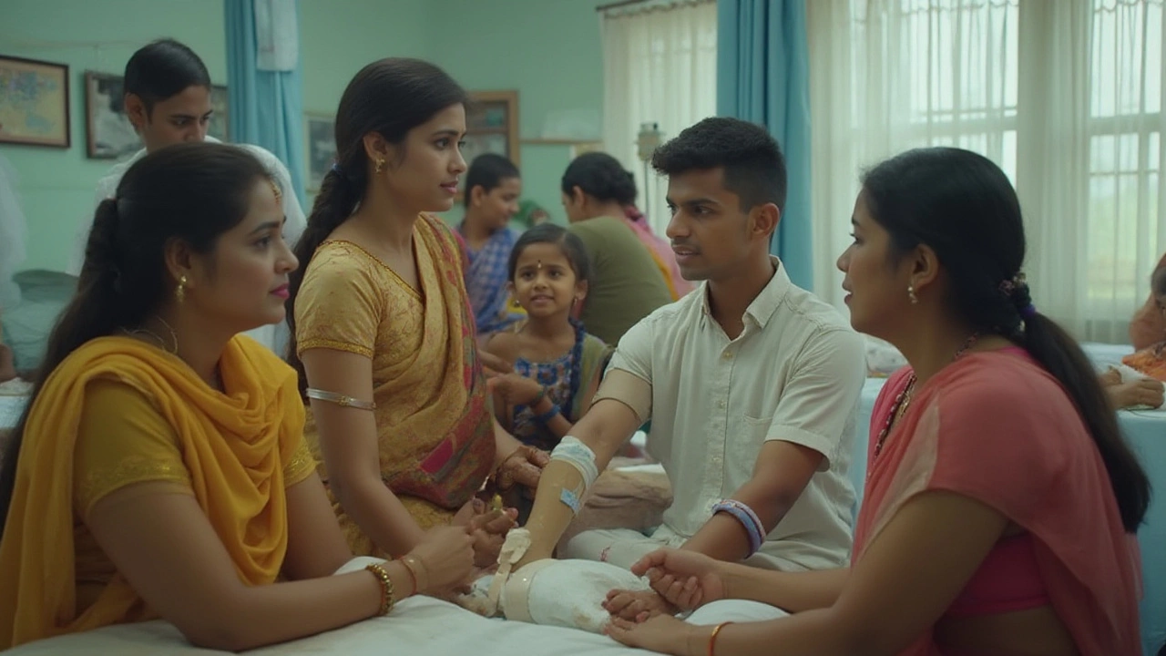
Broken bones and joint problems don’t just hurt—they turn your everyday routine on its head. But when you dig into the world of orthopedics, you quickly notice there’s a playbook behind every cast, splint, and surgical screw. The unsung heroes here? The 4 A’s of orthopedics. These four words—Alignment, Apposition, Apparatus, and Activity—might sound like medical mumbo-jumbo at first glance, but they form the backbone (pun intended) of how orthopedic specialists treat injuries and set patients up for the best possible recovery. Most people don’t know this, but the difference between lifelong stiffness and a full bounce-back sometimes hangs on getting these four pillars right. If you or someone you know has ever hobbled home with a sling or a boot, you’ll want to pay attention.
Alignment: Getting Bones Back on Track
Think about a jigsaw puzzle. If one piece is even a little out of place, the whole picture looks off. That’s pretty much what happens if a bone doesn’t get reset perfectly after a break. Alignment is the first ‘A’—and it’s all about making sure fractured bones are pointing exactly as they should be, just like they were before the injury. This isn’t just about looks; it matters for how your bones carry weight and move again. Imagine your leg bone heals at a weird angle. Every step after that feels wrong or painful. You can’t run or maybe not even walk quite right.
Doctors are obsessed with alignment because if they ignore it, patients can develop what’s called malunion. That’s when bones heal, but in a bad position, causing long-term problems. For example, a 2018 study in the Journal of Orthopaedic Trauma found that nearly 20% of patients with poorly aligned femur fractures ended up with walking difficulties or needed more surgery. That’s a lot of extra pain, time, and money for something that’s fixable early on.
But getting alignment perfect isn’t always easy. Sometimes, bones shift back even after being set—a common problem with wrist and ankle fractures. That’s why you see doctors checking x-rays again and again, sometimes every few days. A little movement can mess things up, so they’ll recast or even repeat surgery if needed. Here’s a tip if you’re recovering from a break: ask for your x-ray results. Get your doctor to show you how the bones line up. Don’t just assume they “look good”—that simple conversation often catches problems early.
Now, let’s talk about some real-world facts. Children’s bones often heal faster, and sometimes, they seem to “self-correct” minor alignment issues. But adults aren’t so lucky—if you’re over 40, bone density drops, and your body can’t fix bad alignment on its own. Staying still during healing might feel boring, but it’s crucial for alignment. If the doctor says “no weight for 6 weeks,” they’re protecting that careful lineup. Otherwise, you could be looking at years of limping or sore joints.
Quick pro tip: Some hospitals now use 3D imaging to check alignment, especially with tricky fractures like the pelvis or jaw. These scans can spot tiny misalignments and save people from big surgeries later. Don’t be shy about asking your orthopedic team what technology they use.
Apposition: Making Sure the Ends Meet
The next step in the healing process is apposition. It sounds technical, but here’s the simple truth—apposition means the broken pieces of bone are touching closely together. There’s no gap or big space between them, and they’re right up against each other so they can knit back naturally. Imagine building a bridge; if the pieces don’t meet in the middle, nobody’s crossing safely.
Now, why does this matter? When there’s a big gap in a fracture, your body can’t lay down new bone tissue fast enough. Instead, it tries to fill the gap with cartilage or fibrous tissue, which is weaker and doesn’t support your weight as well. This can lead to something called non-union, where the bone never fully heals. That often means more surgery and a lot more recovery time. Numbers don’t lie—a recent review in Orthopedic Reviews said that non-union rates can be as high as 15% in severe fractures if apposition isn’t right.
Doctors have a bunch of tricks for getting apposition right. Simple fractures usually just need a careful push and realignment. But if the break is shattered (like in a car crash), they may use metal pins, screws, or even bone grafts to bridge gaps. Ever wonder why surgeons sometimes go back in for a “second look” surgery? That’s often to check and, if needed, improve the apposition in tough fractures. One tip for patients: if your cast or splint feels loose after swelling goes down, get it checked. Movement inside a loose cast can separate bone ends, wrecking early apposition.
Here’s another interesting fact: smokers and people with diabetes or chronic inflammation have a harder time achieving good apposition and healing. Blood flow is a big deal here—without enough new blood vessels growing between bone ends, healing stalls. If you want the best shot at a strong recovery, quit smoking (even temporarily), control diabetes, and eat foods rich in vitamin C and protein. These little changes can boost the body’s ability to lay down the “glue” that holds bone ends together.
Orthopedic teams often track healing progress with x-rays. Look out for words like "callus formation"—that’s your body building a bony bridge across the apposition site. As callus gets denser, you’re moving in the right direction. If it’s not showing up, doctors might recommend electrical bone stimulators or transfer you to a fracture clinic for more aggressive treatment. "Healing is not just waiting, it’s active teamwork between bones, doctors, and patients," says Dr. Michael Simon, Chief of Orthopedic Surgery at a leading trauma center.
“Perfect apposition is where healing starts. If the bones don’t touch, they can’t talk to each other—no matter how good the surgery or cast looks.”
Keep in mind, pets—even active dogs—follow the same rules. Vets use splints and bandages to keep broken legs in apposition, just like with humans. The science holds up across species!
Apparatus and Activity: Tools and Movement in Recovery
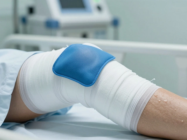
You can set bones perfectly, but if you don’t hold them steady, nature still won’t do its job. Enter Apparatus—the third A. This covers every tool that holds a bone in place while it heals, from simple plaster casts to high-tech fixators and internal plates. The goal here: stability. Without it, any little movement can pull bones out of place, ruining all that careful alignment and apposition work.
The right apparatus depends on the type of fracture and the patient. For a minor finger fracture, a splitting tape does the trick. For a shattered femur, it’s rods and screws. According to a 2023 report in the Journal of Bone & Joint Surgery, over 75% of complex fractures now use some form of internal fixation. These are sturdy metal implants that stay hidden beneath the skin and keep bones in perfect position during daily movement.
Yet, the downside of immobilization? Joints can stiffen quickly and muscles shrink. That’s where Activity, the fourth A, comes in. Activity means carefully reintroducing movement once stability is secure. Too soon, and bones may not unite; too late, and you risk permanent stiffness. Striking the right balance is almost an art. Physical therapists make a huge impact here—they teach specific exercises that rebuild strength without endangering the new bone. For instance, after an elbow fracture, doing “table slides” with a towel helps get movement back without straining the healing spot.
Check out these tips and facts:
- Most casts should stay dry. Water can weaken plaster or cause skin sores. If you must shower, cover your cast with a watertight bag.
- Some removable braces are designed for nightly wear but can be taken off during controlled activity—always ask before removing yours.
- Early gentle movements (like wiggling your toes with an ankle cast) keep blood flowing and reduce swelling. But never try weight-bearing before your doc gives the nod.
- Physical therapy can start as soon as the fracture is stable, sometimes within a week for minor breaks.
- A recent patient satisfaction survey from NHS England found that patients who started guided rehab within two weeks of injury had a 50% greater chance of regaining full range of motion compared to those who waited six weeks or more.
Here’s a look at how apparatus and activity choices break down by fracture type:
| Fracture Site | Apparatus Used | Recommended Activity |
|---|---|---|
| Wrist | Plaster cast, removable splint | Finger exercises early; full wrist movement after 4-6 weeks |
| Femur | Internal rod, external fixator | Early walking with crutches; full weight after 8-10 weeks |
| Ankle | Air-cast boot, surgical screws | Non-weight initially; gradual load-bearing after healing confirmed |
| Shoulder | Sling, sometimes plate/screws | Pendulum swings day 2-3; resistance training after 4-6 weeks |
Don’t forget: The time you spend wearing the apparatus matters. Most healing happens in the first 6 weeks. But bones keep remodeling for months afterward. Skipping out early—ditching your boot for a hike or slacking on exercises—has sent plenty of folks right back to hospital beds. If a doctor or physio gives you a timeline, follow it even if things “feel okay” halfway through.
If your device causes pain, numbness, or swells up badly, head back to your ortho team. Those are warning signs that the apparatus may be on too tight or in the wrong spot—ignoring them can cost you healing time or even lead to nerve damage.
Bottom line: The 4 A’s of orthopedics aren’t just medical buzzwords. They’re a daily checklist used in trauma rooms, clinics, and even on the sports field. If you understand why they matter, you’re way more likely to avoid the classic horror stories—stiff limbs, repeat surgeries, endless pain—that spoil recovery for so many active people and weekend warriors. Next time you or a friend breaks something, remember: alignment gets you straight, apposition gets you close, apparatus holds it all together, and activity brings you back to action. Funny how four simple A-words can spell out the whole secret to moving well after an injury.